GFAP Protein – Prot-p-GFAP
Porcine (Pig) GFAP protein

$605.00 CAD
0 in stock (expect 7-21 days for delivery)
0 in stock (expect 7-21 days for delivery)
GFAP Protein – Prot-p-GFAP
GFAP Purified Protein : Glial Fibrillary Acidic Protein (GFAP) is a major protein of the nervous system and is localized in astrocytes, stem cells, Bergmann glia and non-myelinating Schwann cells. It may also be found in retinal Mueller cells in pathological states, and the levels of the protein generally increase in damage and disease states. GFAP assembles to form 10nm or intermediate filaments in the cytoplasm, and these filaments appear to have an important structural role in the cell.
Characteristics of Porcine (Pig) GFAP protein
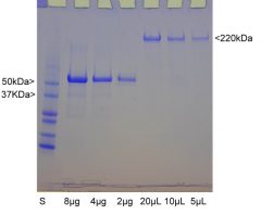
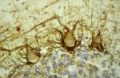
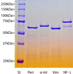
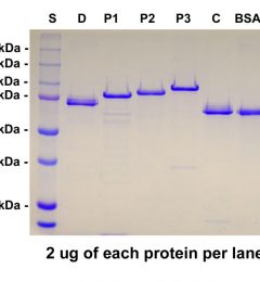
Background: Glial Fibrillary Acidic Protein (GFAP) was discovered by Amico Bignami and coworkers as a major fibrous protein of multiple sclerosis plaques (2). It was subsequently found to be a member of the 10nm or intermediate filament protein family, specifically the intermediate filament protein family Class III, which also includes peripherin, desmin and vimentin. The one mammalian GFAP gene produces several transcripts producing several different possible protein products (1). In pigs there are two major protein products, one of 428 amino acids and a second including a 40 amino acid insert in the C-terminal region. The two proteins are of about equal abundance and run on gels at about 50kDa and 54kDa, so purified pig GFAP therefore presents as two closely spaced bands as shown in the gel image. The larger protein is in Genbank here and the lower here. Both forms bind most available GFAP antibodies including our mouse monoclonal MCA-5C10 and our rabbit and chicken polyclonals, RPCA-GFAP and CPCA-GFAP. GFAP is strongly and specifically expressed in astrocytes and certain other astroglia in the central nervous system, in satellite cells in peripheral ganglia, and in non-myelinating Schwann cells in peripheral nerves. In many damage and disease states GFAP expression is heavily upregulated in astrocytes. In addition neural stem cells frequently strongly express GFAP. Antibodies to GFAP are therefore very useful as markers of astrocytic cells and neural stem cells. In addition many types of brain tumor, presumably derived from astrocytic cells, heavily express GFAP. Alexander’s disease was recently shown to be caused by point mutations in the protein coding region of the GFAP gene (3). All forms of Alexander disease are characterized by the presence of Rosenthal fibers, which are GFAP containing cytoplasmic inclusions found in astrocytes. There has been considerable recent interest in GFAP due to potential use as a damage and degeneration biomarker, since it can be detected in blood and CSF following various kinds of CNS damage and disease states (5). This GFAP preparation is an excellent protein standard for such experiments. Since the human and rodent proteins are somewhat different in primary sequence from each other and from the pig protein, we have also generated recombinant forms of these specifically Prot-r-GFAP and Prot-r-GFAP-rat. The HGNC name for this protein is GFAP.
Encor GFAP Protein – Prot-p-GFAP
Formats available : 50 ug, 100 ug
Link to GFAP Protein – Prot-p-GFAP on Encor’s website
References :
1. Leung, C. L. and Liem, R. K. H. Isolation of intermediate filaments. Curr. Prot. Cell Biol. 3:Unit 3.23 doi: 10.1002/0471143030.cb0323s31 (2006).
2. Bignami A, Eng LF, Dahl D, Uyeda CT. Localization of the glial fibrillary acidic protein in astrocytes by immunofluorescence. Brain Res. 43:429-35 1972.
3. Brenner M, Johnson AB, Boespflug-Tanguy O, Rodriguez D, Goldman JE and Messing A. Mutations in GFAP, encoding glial fibrillary acidic protein, are associated with Alexander disease. Nat Genet 27:117-20 2001
4. Liem RKH, Yen SH, Salomon GD and Shelanski ML. Intermediate filaments in nervous tissues. J Cell Biol 79:637-745 (1978).
5. Schiff L1, Hadker N, Weiser S, Rausch C. A literature review of the feasibility of glial fibrillary acidic protein as a biomarker for stroke and traumatic brain injury. Mol Diagn Ther 16:79-92 (2012).
Additional information
| Format | 50 ug |
|---|

Reviews