pNF-H ELISA Kit Version 2 – ELISA-pNF-H-V2
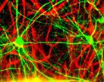
Neurofilament subunit NF-H is one of the three so-called neurofilament triplet proteins which are major structural components of neurons. These proteins are particularly heavily concentrated in the axons of large projection neurons, where they clearly occupy the majority of the cytoplasm. The NF-H molecule has a highly unusual protein sequence which includes a highly repetitive region made up of slight variants of the 6-8 amino acid sequences based on the peptide lysine-serine-proline (KSP in the single letter code). There are about 50 of these tandemly repeated peptides in known mammalian NF-H sequences, the exact number being a little variable in different species. Interestingly, the serine residues are not phosphorylated in dendritic and perikaryal neurofilaments but are heavily phosphorylated in axonal neurofilaments, so that antibodies which recognize the KSP phosphorylated from NF-H, which we refer to as pNF-H, are excellent markers of axons (1,2). We have developed a sensitive ELISA kit to accurately quantify the level of expression of this protein in a variety of experimental situations. Since NF-H is expressed later in development, and becomes heavily phosphorylated later than the other two triplet proteins this kit can be used to monitor neuronal maturation. Neurofilament expression is up regulated in a variety of damage and disease states, and this kit can be used to accurately quantify this up regulation. We have also recently found that this kit can detect pNF-H in the sera of animals with spinal cord and brain lesions, suggesting the exciting possibility that pNF-H is a useful biomarker of neuronal and more specifically axonal injury or degeneration (3). Further studies show that this assay can detect informative levels of pNF-H in both the CSF and blood of humans suffering from a variety of neurological disorders (4,5,6). In fact, there are several reasons to think that pNF-H might be an unusually useful and informative biomarker of axonal injury and degeneration. Firstly, pNF-H is an abundant component of axons as noted above, so it should be released in relatively large quantity following axonal damage and degeneration. Secondly, it is unusually resistant to proteolysis, apparently partly due to the heavy phosphorylation (7,8), meaning that it is likely to be relatively long lived on release into CSF and blood, aiding its detection. Thirdly, the unusual multiepitope nature of the phosphorylation sites coupled with their great immunogenicity means that pNF-H can be captured and detected with unusual avidity by appropriate immunoreagents. Finally, pNF-H detection reflects axonal injury and much recent evidence suggests that a variety of damage and disease states can be regarded as the result of axonal injury (e.g. 9,10). The unique geometry of neurons renders the axons particularly sensitive to mechanical injury or local loss of oxygen or metabolic substrates, and our assay can determine how much of this axonal injury has occurred. This kit was recently used to show that pNF-H levels were greatly elevated in the CSF of patients with subacute sclerosing panencephalitis, and that the levels detected correlated well with the state of progression of the disease (11).
Characteristics of the pNF-H ELISA Kit Version 2
The pNF-H ELISA Kit Version 1 can be used for the detection of pNF-H in plasma, serum, CSF and tissue extracts.
To develop the pNF-H ELISA Kit Version 2, we purified a monoclonal antibody to pNF-H, MCA-AH1 and applied 50 ng of this pure protein to each well of the ELISA dish. This is an unusually high affinity and specificity antibody which was selected out of several candidates for its ability to pull pNF-H out of concentrated protein solutions (12). To detect the binding of pNF-H to this monoclonal antibody we made use of a second pNF-H monoclonal antibody, clone MCA-NAP4. This antibody was directly labelled with horse radish peroxidase, providing an assay that is quicker and more convenient than our previous assay.
The kit contains one standard ELISA 96 well plate coated with the chicken antibody which has been blocked and so it is ready to use. The plate can be separated into 8 x 12 well strips. The kit includes enough of the rabbit anti pNF-H detection antibody and a pure pNF-H standard. Also included are a goat anti-rabbit alkaline phosphatase conjugate, p-Nitrophenol phosphatase substrate and buffer concentrates. We also include detailed instructions.
Reactivity: Human, horse, cow, pig, chicken, rat, mouse
Immunogen: Phosphorylated axonal NF-H (pNF-H)
Isotype: Mouse IgG1
Mass of detected protein: 110 kDa
Application and sample type: ELISA of blood, CSF and tissue extracts.
Storage: Shipped on ice. Store at 4°C.
Encor pNF-H ELISA Kit Version 2 – ELISA-pNF-H-V2
Formats available : 96 rx
Link topNF-H Version 2 ELISA Kit – ELISA-pNF-H-V2 on Encor’s website
References :
1. Sternberger LA, Sternberger NH. Monoclonal antibodies distinguish phosphorylated and nonphosphorylated forms of neurofilaments in situ. PNAS 80:6126-6130 (1983).
2. Lee VM, Otvos L Jr, Carden MJ, Hollosi M, Dietzschold B, Lazzarini RA. Identification of the major multiphosphorylation site in mammalian neurofilaments. PNAS 85:1998-2002 (1998).
3. Shaw G, Yang C, Ellis R, Anderson K, Parker Mickle J, Scheff S, Pike B, Anderson DK and Howland DR. Hyperphosphorylated neurofilament NF-H is a serum biomarker of axonal injury. Biochem Biophys Res Commun. 336:1268-1277 (2005).
4. Petzold, A. and Shaw, G. Comparison of two ELISA methods for measuring levels of the phosphorylated neurofilament heavy chain. J Immunol Methods 319:34-40 (2007).
5. Siman, R. et al. Biomarker evidence for mild central nervous system injury after surgically-induced circulation arrest. Brain Res. [Epub ahead of print] (2008).
6. Lewis, S. et al. Detection of phosphorylated NF-H in the cerebrospinal fluid and blood of aneurysmal subarachnoid hemorrhage patients. J Cereb Blood Flow Metab. Epub Mar 5 (2008).
7. Schlaepfer WW, Lee C, Lee VM, and Zimmerman UJ. An immunoblot study of neurofilament degradation in situ and during calcium-activated proteolysis, J. Neurochem. 44:502–509 (1985).
8. Pant HC. Dephosphorylation of neurofilament proteins enhances their susceptibility to degradation by calpain.
Biochem J. 256:665-8 (1988).
9. Stys PK. General mechanism of axonal damage and its prevention. J. Neurol. Sci. 233:3-13 (2005).
10. Buki A and Povlishock JT. All roads lead to disconnection? – Traumatic axonal injury revisited.
Acta Neurochir (Wien). 148:181-93 (2005).
11. Matsushige T, Ichiyama T, Anlar B, Tohyama J, Nomura K, Yamashita Y, Furukawa S. CSF neurofilament and soluble TNF receptor 1 levels in subacute sclerosing panencephalitis. J Neuroimmunology [Epub ahead of print] (2008).
12. Boylan, K., Yang, C., Crook, J., Overstreet, K., Heckman, M., Wang, Y., Borchelt, D. and Shaw, G. Immunoreactivity of the phosphorylated axonal neurofilament H subunit (pNF-H) in blood of ALS model rodents and ALS patients: Evaluation of blood pNF-H as a potential ALS biomarker. J. Neurochem 111:1182-1191 (2009).
Additional information
| Format | 96 rx |
|---|


Reviews