Lamp1 Antibody – MCA-6E2
- Clonality : Mouse Monoclonal
- Applications : WB | IF/ICC | IHC
- Reactivity : Human | Horse | Cow | Pig | Chicken | Rat | Mouse


$338.00 – $1,810.00 CAD
0 in stock (expect 7-21 days for delivery)
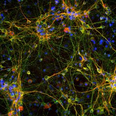
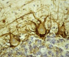
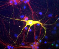
Encor Mouse Monoclonal to Lysosomal Associated Membrane Protein 1 (LAMP1, a.k.a. CD107a, LGP120 and LAMPA), LAMP1
As the name suggests, LAMP1 is a protein primarily associated with the lysosomal membrane. Antibodies to LAMP1 are therefore excellent markers of lysosomes in mammalian cells, though some LAMP1 may also be seen on late endosomes and on the plasma membrane. In a typical cell LAMP1 is associated with spherical vesicles located next to the nucleus and the microtubule organizing center (1). The protein is also known as CD107a, lysosomal associated membrane glycoprotein 1, LGP120 and LAMPA, as the protein was independently discovered and named by several different labs. CD is an abbreviation for “Cluster of Differentiation” and refers to a protocol for the naming of proteins and other surface markers of human leukocytes defined by binding of specific monoclonal antibodies.
LAMP1 is found on the cell surface of lymphocytes undergoing degranulation, a process in which cytoplasmic vesicles fuse with the plasma membrane, and this phenomena resulted in discovery of LAMP1 as a CD protein.
The LAMP1 protein has a large N-terminal region which is inside the lysosome, hence topologically external to the cell, which is often referred to as the lumenal domain (2). The lumenal domain consists of two homologous globular segments separated by a proline rich sequence. Next there is a single membrane spanning domain and a short 11 amino acid C-terminal cytoplasmic tail. This tail region contains, at the extreme C-terminus, a so-called YXXI motif which is responsible for the sorting of the intact molecule to the endosome and lysosome, where Y = tyrosine, I = isoleucine and X = almost any amino acid (3). This motif is found in several other lysosomal proteins, where it functions in the same way. There are 417 amino acids in the human LAMP1 molecule, giving a native molecular weight of 44.8 kDa. However, the N-terminal lumenal segment of LAMP1 is very heavily and variably glycosylated due to the presence of 18 N-linked glycosylation sites, so that on SDS-PAGE and on Western blots the protein runs as a diffuse band at 90-120 kDa. The HGNC name for this protein is LAMP1.
HGNC name(s) : LAMP1
Host : Mouse
Clonality : Monoclonal
ID : EnCor Biotechnology Lamp1 6E2
Reactivity : Human | Horse | Cow | Pig | Chicken | Rat | Mouse
Isotype : IgG1
Conjugation : none
Immunogen : Recombinant full length human
Mass of detected protein : ~90 and 120 kDa
Uniprot ID : P11279
KGNC name : LAMP1
RRID # : AB_2572343
Purification : Affinity purified at 1 mg/mL
Storage : Shipped on ice. Store at 4°C. For long term storage, leave frozen at -20°C. Avoid freeze / thaw cycles.
Validated applications : WB | IF/ICC | IHC
Suggested Dilutions:
WB: 1:5 000. ICC/IF and IHC: 1:500.
References :
1. Matteoni, R. and Kreiss, T. E. Translocation and clustering of endosomes and lysosomes depends on microtubules. J. Cell Biol. 105:1253-1265 (1987).
2. Howe CL, Granger BL, Hull M, Green SA, Gabel CA, Helenius A, Mellman I. Derived protein sequence, oligosaccharides, and membrane insertion of the 120-kDa lysosomal membrane glycoprotein (lgp120): identification of a highly conserved family of lysosomal membrane glycoproteins. Proc Natl Acad Sci U S A. 85:7577-81 (1988).
3. Rohrer J, Schweizer A, Russell D, Kornfeld S. The targeting of Lamp1 to lysosomes is dependent on the spacing of its cytoplasmic tail tyrosine sorting motif relative to the membrane. J Cell Biol. 132:565-76 (1996).
Additional information
| Format | 50 ul, 100 ul, 500 ul |
|---|---|
| Supplier | |
| Host | Mouse |
| Clonality | Monoclonal |
| Conjugation | None |
Ask a question about Lamp1 Antibody – MCA-6E2
Related products
-

Microtubule Associated Protein tau, MAPT Antibody – CPCA-Tau
$338.00 – $1,810.00 CAD -

Neurofilament NF-H Antibody – MCA-NAP4
$338.00 – $1,810.00 CAD -
GAP43 Antibody – CPCA-GAP43
$338.00 – $1,810.00 CAD -
Peripherin Antibody – CPCA-Peri
$338.00 – $1,810.00 CAD -
Vimentin Antibody – CPCA-Vim
$338.00 – $1,810.00 CAD -

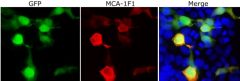
Green Fluorescent Protein, GFP Antibody – MCA-1F1
$338.00 – $1,810.00 CAD


Reviews