Coronin 1a Antibody – RPCA-Cor1a
- Clonality : Rabbit Polyclonal
- Applications : WB | IF/ICC | IHC
- Reactivity : Human | Horse | Cow | Pig | Chicken | Rat | Mouse


$338.00 – $1,810.00 CAD
0 in stock (expect 7-21 days for delivery)
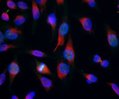
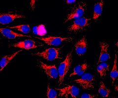
Encor Rabbit polyclonal to Coronin 1a
Coronin was originally discovered in Dictyostelium, where it was found to be involved in the chemotactic response of these amoeboid cells (1,2). The name derives from the fact that the protein is localized at the leading edge or crown of these highly motile cells. The name derives from Corona, which is Latin for crown.
Coronin homologues have been found in yeast, C. elegans, Drosophila and many other species, and a family of them are known in mammals. All coronins belong to the WD40 or WD family of proteins, the prototype which is the β subunit of trimeric G-proteins. The β subunit proteins all have a beautiful conserved wheel-like seven bladed β-propeller structure, with each blade being formed by four β strands generated by one of the WD sequence repeats. The G protein β subunits are believed to function as general purpose binding adapters, mediating numerous regulatory binding interactions between G proteins, G protein coupled receptors and G protein effectors.
Although sequence analysis suggested that coronin 1a only possesses 5 of the these WD repeats, recent structural studies show that, like the G protein β-subunits, there are seven β-propellers, generating a compact 7 bladed propeller (3). Coronins appear to be particularly involved in binding to actin, actin associated proteins, tubulin and phospholipase C and have been implicated in the mechanisms of chemotaxis and phagocytosis.
In mammals, there are at least five major coronin proteins, named coronins 1 to 5 in one nomenclature. Another nomenclature divides these five proteins in coronins 1a and 1b, 2a, 2b and 2c (see the Human Genome Organization Gene Nomenclature Committee link for this family). The various coronin proteins have several other alternate names, since they were discovered independently by several different groups. The mammalian coronin family members are abundant components of eukaryotic cells, and each type has a restricted cell type specific expression pattern. Coronin 1A is the mammalian coronin most similar in protein sequence to the Dictyostelium protein and is found exclusively in hematopoetic lineage cells such as lymphocytes, macrophages and neutrophils.
Therefore, RPCA-Cor1a is an excellent marker of cells of this lineage and can also be used to study the leading edges particularly of neutrophils. Since the only hematopoetic cells found within the central nervous system are microglia, this antibody is also an excellent marker of this important cell type (4). Microglia are numerically fairly minor components of the nervous system, but microglial activation is seen in response to a wide variety of damage and disease states, including ALS, Alzheimer’s disease and responses to brain tumors. Since coronin 1a is a constitutive component of microglia, the coronin 1a antibody can be used to study both quiescent and activated microglia.
The HGNC name for this protein is CORO1A.
HGNC name(s) : CORO1A
Host : Rabbit
Clonality : Polyclonal
ID : EnCor Biotechnology Coronin 1a Cor1a
Reactivity : Human | Horse | Cow | Pig | Chicken | Rat | Mouse
Isotype : IgG
Conjugation : none
Immunogen : Recombinant full length human
Mass of detected protein : 57 kDa
Uniprot ID : P31146
KGNC name : CORO1A
RRID # : AB_2229659
Purification : Serum
Storage : Shipped on ice. Store at 4°C. For long term storage, leave frozen at -20°C. Avoid freeze / thaw cycles.
Validated applications : WB | IF/ICC | IHC
Suggested Dilutions:
WB: 1:20 000, IF/ICC: 1:500-1:1 000, ABC: 1:5 000
References :
1. de Hostos, E. The coronin family of actin-associated proteins. Trends in Cell Biology 9:345-350 (1999).
2. Rybakin, V. and Clemen, C. S. Coronin proteins as multifunctional regulators of the cytoskeleton and membrane trafficking. BioEssays. 27:625-632 (2005).
3. Appleton, B. A., Wu, P and Weisman, C. The crystal structure of murine coronin-1: a regulator of actin cytoskeletal dynamics in lymphocytes. Structure 14:87-96 (2006).
4. Ahmed Z, Shaw G, Sharma VP, Yang C, McGowan E, Dickson DW. The Actin Binding Proteins Coronin-1a and IBA-1 Are Effective Microglial Markers for Immunohistochemistry J. Histochem. Cytochem. 2007 (Epub).
Additional information
| Format | 50 ul, 100 ul, 500 ul |
|---|---|
| Supplier | |
| Host | Rabbit |
| Clonality | Polyclonal |
| Conjugation | None |
Ask a question about Coronin 1a Antibody – RPCA-Cor1a
Related products
-

Aurora B Kinase Antibody – MCA-6G2
$338.00 – $1,810.00 CAD -
Vimentin Antibody – CPCA-Vim
$338.00 – $1,810.00 CAD -

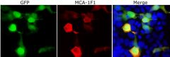
Green Fluorescent Protein, GFP Antibody – MCA-1F1
$338.00 – $1,810.00 CAD -

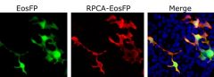
Green-Red photoconvertible fluorescent protein EosFP Antibody – RPCA-EosFP
$338.00 – $1,810.00 CAD -
Heat shock protein 60, HSP60 Antibody – MCA-1C7
$338.00 – $1,810.00 CAD -
Pin1 Antibody – CPCA-Pin1
$338.00 – $1,810.00 CAD

Reviews